Peptides and Hip Pain: Exploring Research on Bursitis, Tendinopathy, and Joint Recovery
Hip pain from bursitis, gluteal tendinopathy, or labral pathology can be debilitating. This article examines how BPC-157 and TB-500 research relates to the specific tissue biology of common hip conditions.
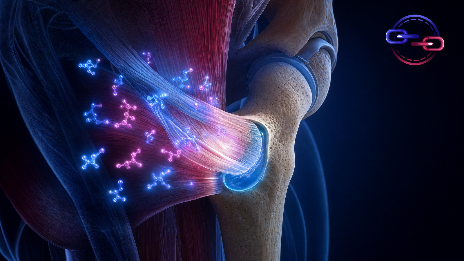
Hip pain is one of the most common musculoskeletal complaints, particularly among active adults and aging populations. What was traditionally diagnosed as "trochanteric bursitis" is now understood to be primarily gluteal tendinopathy — a degenerative process affecting the tendons that attach the gluteal muscles to the greater trochanter of the femur. This shift in understanding has significant implications for how we think about treatment approaches, including the potential relevance of tissue repair peptides.
Greater Trochanteric Pain Syndrome: Beyond Bursitis
For decades, lateral hip pain was attributed almost exclusively to inflammation of the trochanteric bursa. However, MRI and surgical studies have revealed that the majority of cases involve degeneration and tearing of the gluteus medius and gluteus minimus tendons at their insertion on the greater trochanter. The condition is now more accurately termed Greater Trochanteric Pain Syndrome (GTPS), which encompasses gluteal tendinopathy, bursitis, and iliotibial band friction.
This is significant because tendinopathy and bursitis require fundamentally different approaches. Bursitis is inflammatory — it responds to anti-inflammatory interventions. Tendinopathy is degenerative — it requires tissue repair strategies. Understanding which pathology predominates guides both conventional treatment and research directions.
Gluteal Tendinopathy: The Rotator Cuff of the Hip
Orthopedic researchers increasingly describe the gluteal tendons as "the rotator cuff of the hip" — and the analogy is remarkably apt. Like the rotator cuff, the gluteal tendons operate through a narrow anatomical space, experience complex multi-directional forces, have regions of limited blood supply, and undergo progressive degenerative changes that are often bilateral.
The gluteus medius tendon, in particular, shows pathological changes nearly identical to those seen in supraspinatus tendinopathy: collagen disorganization, mucoid degeneration, neovascularization, and partial-thickness tearing. These changes reduce the tendon's mechanical capacity and contribute to the characteristic lateral hip pain that worsens with walking, climbing stairs, and lying on the affected side.
BPC-157 and Hip Tendon Biology
BPC-157's published tendon research applies directly to gluteal tendinopathy through the same mechanisms relevant to other tendon conditions. VEGF-mediated angiogenesis addresses the hypovascular zones within the gluteal tendon insertion. Growth factor receptor upregulation may enhance the tissue's capacity to respond to repair signals. And the FAK-paxillin pathway activation promotes the fibroblast migration and adhesion necessary for collagen matrix production.
The NO system modulation is particularly interesting for hip conditions because the greater trochanteric region receives blood supply from multiple sources (superior gluteal artery, lateral circumflex femoral artery), and NO signaling plays a key role in regulating blood flow distribution to the tendon insertion zone.
TB-500 and Hip Recovery
TB-500's cell migration promotion is relevant because gluteal tendon tears, like rotator cuff tears, face healing challenges related to the size of the defect and the distance repair cells must travel. In partial-thickness tears, the remaining tendon tissue must recruit sufficient fibroblasts to produce new collagen and restore mechanical integrity. TB-500's actin-regulation mechanism facilitates this cellular recruitment.
The anti-inflammatory properties of TB-500 address the bursitis component of GTPS. Even when tendinopathy is the primary pathology, secondary bursitis often develops as the bursa becomes irritated by the dysfunctional tendon mechanics. Modulating the inflammatory environment may help create more favorable conditions for tendon healing while also reducing pain from bursal inflammation.
Hip Labral Pathology
The acetabular labrum — a ring of fibrocartilage that deepens the hip socket and improves joint stability — is another common source of hip pain. Labral tears can result from trauma, repetitive motion, or structural variations like femoroacetabular impingement (FAI). Like other fibrocartilage structures, the labrum has limited blood supply peripherally and essentially no blood supply centrally, making healing difficult.
Published BPC-157 research on angiogenesis and connective tissue repair has theoretical relevance to labral healing, though no specific labral studies have been published. TB-500's promotion of cell migration could be relevant for facilitating repair cell access to the avascular central labral zone.
Conventional Evidence-Based Treatment
Evidence-supported approaches for GTPS include load management and activity modification to reduce compressive forces on the gluteal tendons, progressive gluteal strengthening with attention to the gluteus medius and minimus, avoidance of positions that compress the tendons (crossing legs, prolonged side-lying on the affected hip), addressing contributing factors such as weak hip abductors and ITB tightness, and medical evaluation for cases not responding to conservative management within 3-6 months.